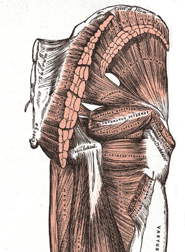
 Zone 01Gray’s Anatomy, 1918
Hips, Gluteal Complex & Pelvic Girdle
Gluteus medius/minimus, piriformis, deep hip rotators & acetabular labral capsular restrictions.
Included in Practical Lab
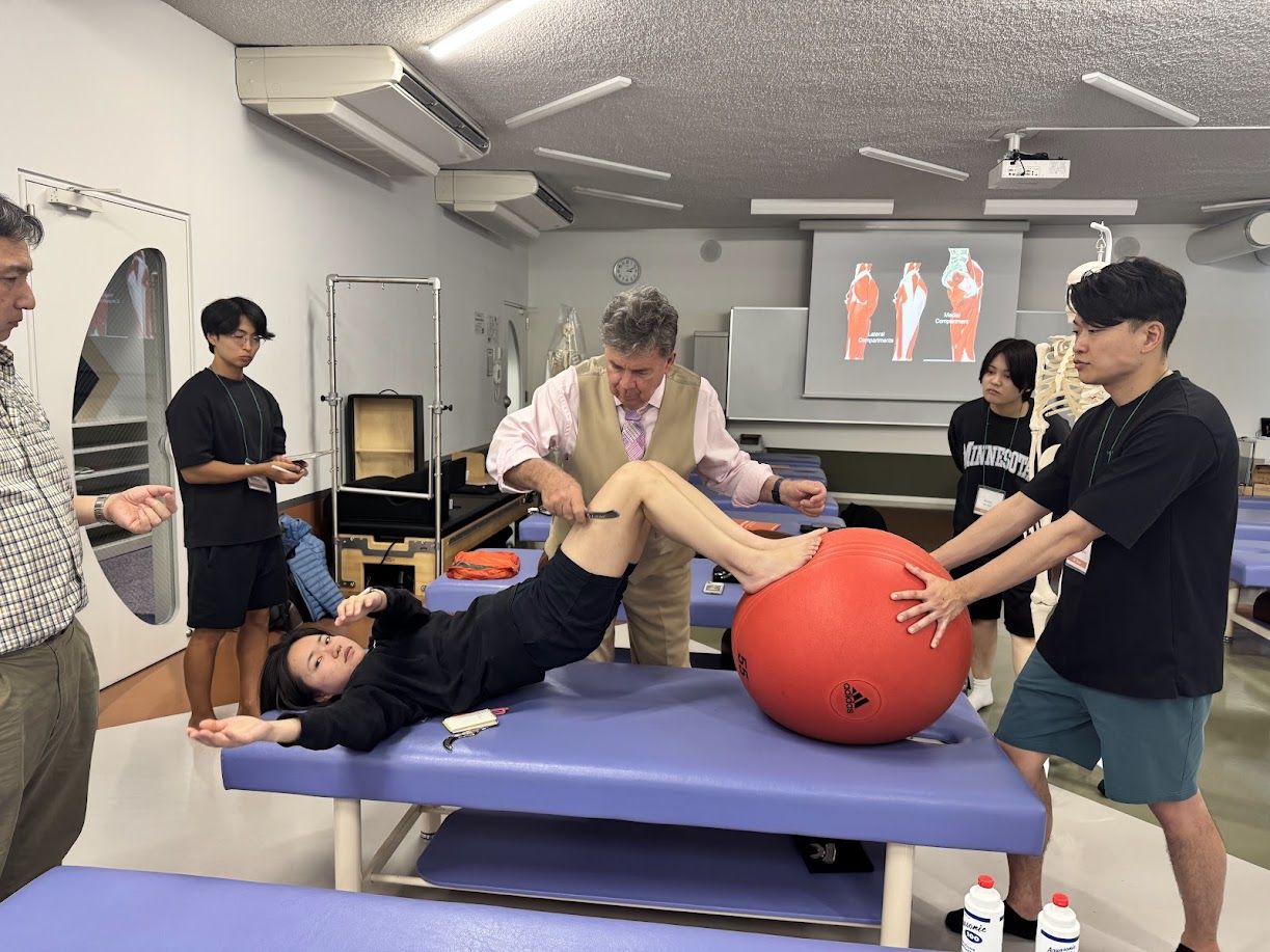
Advanced instruction in soft-tissue restrictions, passive movement assessment, and precise manual manipulation of the lower kinetic chain.
Hands-on clinical positioning for hip, knee, ankle, foot, and sciatic neurovascular releases.
DRT Lower Extremity provides advanced instruction in the assessment and treatment of soft-tissue restrictions involving the feet, ankles, lower legs, knees, thighs, hips, and surrounding structures.
DRT Lower Extremity provides advanced instruction in the assessment and treatment of soft-tissue restrictions involving the feet, ankles, lower legs, knees, thighs, hips, and surrounding structures. Attendees review the relevant muscles, tendons, ligaments, fascial planes, connective tissues, and neurovascular structures while learning the appropriate application of Dynamic Release Techniques.
Anatomy & PalpationBecause the patient may be unconscious under anesthesia, all positioning and movement are performed passively by the treating physician and trained assistant. The patient does not actively contract, resist, or control the movement. This allows the physician to assess tissue tension, joint restriction, adhesions, and movement barriers while applying precise Passive Release Techniques before progressing to osseous manipulation when clinically appropriate.
Anesthesia SettingThe course emphasizes controlled lower-extremity positioning, physician-assisted movement, tissue-specific contact, patient safety, communication with the anesthesia team, and the proper sequence of soft-tissue treatment and manipulation.
Safety & SequencingIn-depth review of hips, thighs, knees, lower legs, ankles, and feet muscles, tendons, fascia, and neurovascular entrapments.
Physician-assisted lower limb mobilization without active patient resistance under unconscious or passive anesthesia conditions.
Seamless coordination with anesthesia professionals to ensure proper patient positioning, monitoring, and soft-tissue sequence.
Each zone is covered in the online anatomy review and practiced hands-on in the lab.
Zone 01Gray’s Anatomy, 1918Gluteus medius/minimus, piriformis, deep hip rotators & acetabular labral capsular restrictions.
Included in Practical Lab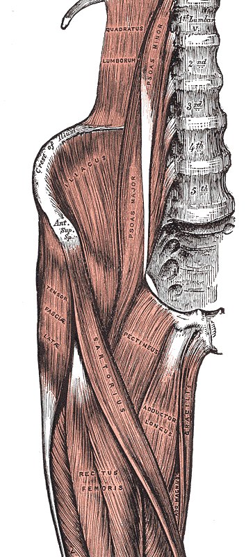 Zone 02Gray’s Anatomy, 1918
Rectus femoris, vastus musculature, biceps femoris, semitendinosus & iliotibial (IT) band fascia.
Included in Practical Lab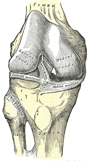 Zone 03Gray’s Anatomy, 1918
Patellofemoral tracking, collateral & cruciate ligamentous tension, popliteus & meniscal margins.
Included in Practical Lab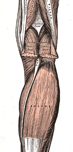 Zone 04Gray’s Anatomy, 1918
Anterior/posterior compartments, tibialis anterior, gastrocnemius, soleus & interosseous membrane.
Included in Practical Lab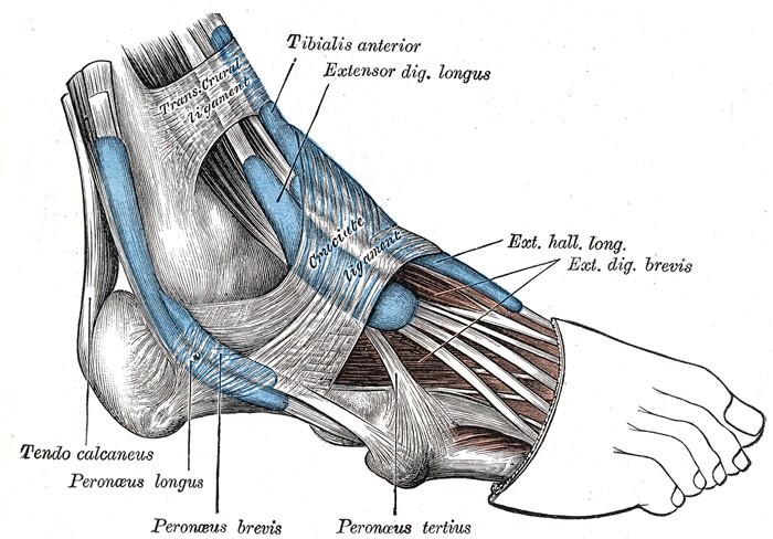 Zone 05Gray’s Anatomy, 1918
Talocrural & subtalar joint mobility, Achilles tendon insertion, inferior/superior peroneal retinaculum.
Included in Practical Lab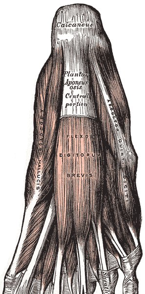 Zone 06Gray’s Anatomy, 1918
Plantar aponeurosis, flexor digitorum brevis, tarsal tunnel nerve pathways & intrinsic foot arches.
Included in Practical LabAnatomical plates: Henry Gray, Anatomy of the Human Body (1918), public domain, via Wikimedia Commons.
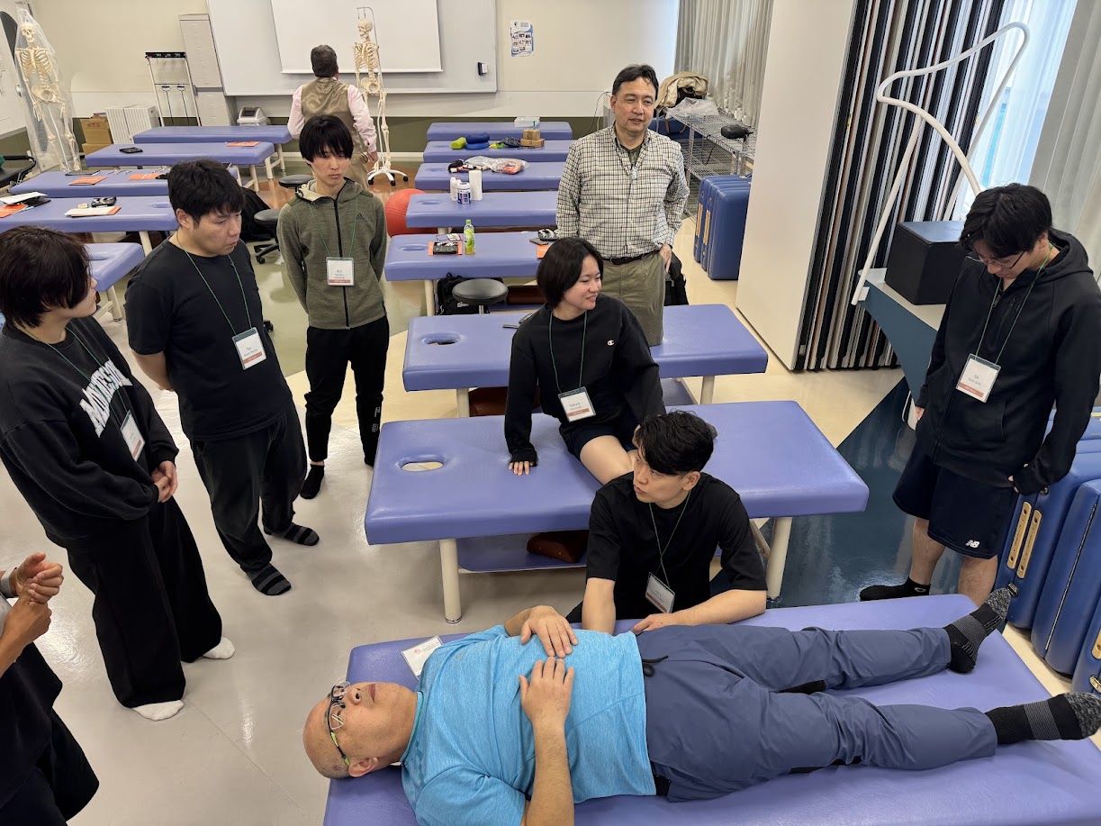
Attendees receive personal guidance from certified DRT faculty, practicing lower extremity lever angles, tissue tension palpation, and safe positioning maneuvers on live subjects in first-class seminar venues worldwide.
Explore the other regions
Join leading healthcare professionals in our upcoming destination seminars and add DRT Lower Extremity to your clinical skillset.